The circulatory system of earthworm is closed type, as the blood flows only thorough the blood vessels. The blood vascular system of Pheretima posthuma consists of an elaborate system of blood vessels through which blood flows continuously. The blood of earthworm is red in color due to the presence of hemoglobin dissolved in the plasma unlike other vertebrates where hemoglobin is present in the red blood cells. The cells suspended in the plasma are transparent and colorless.
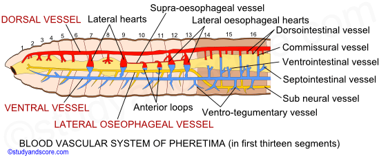
The blood vessels and their arrangement in first thirteen segments
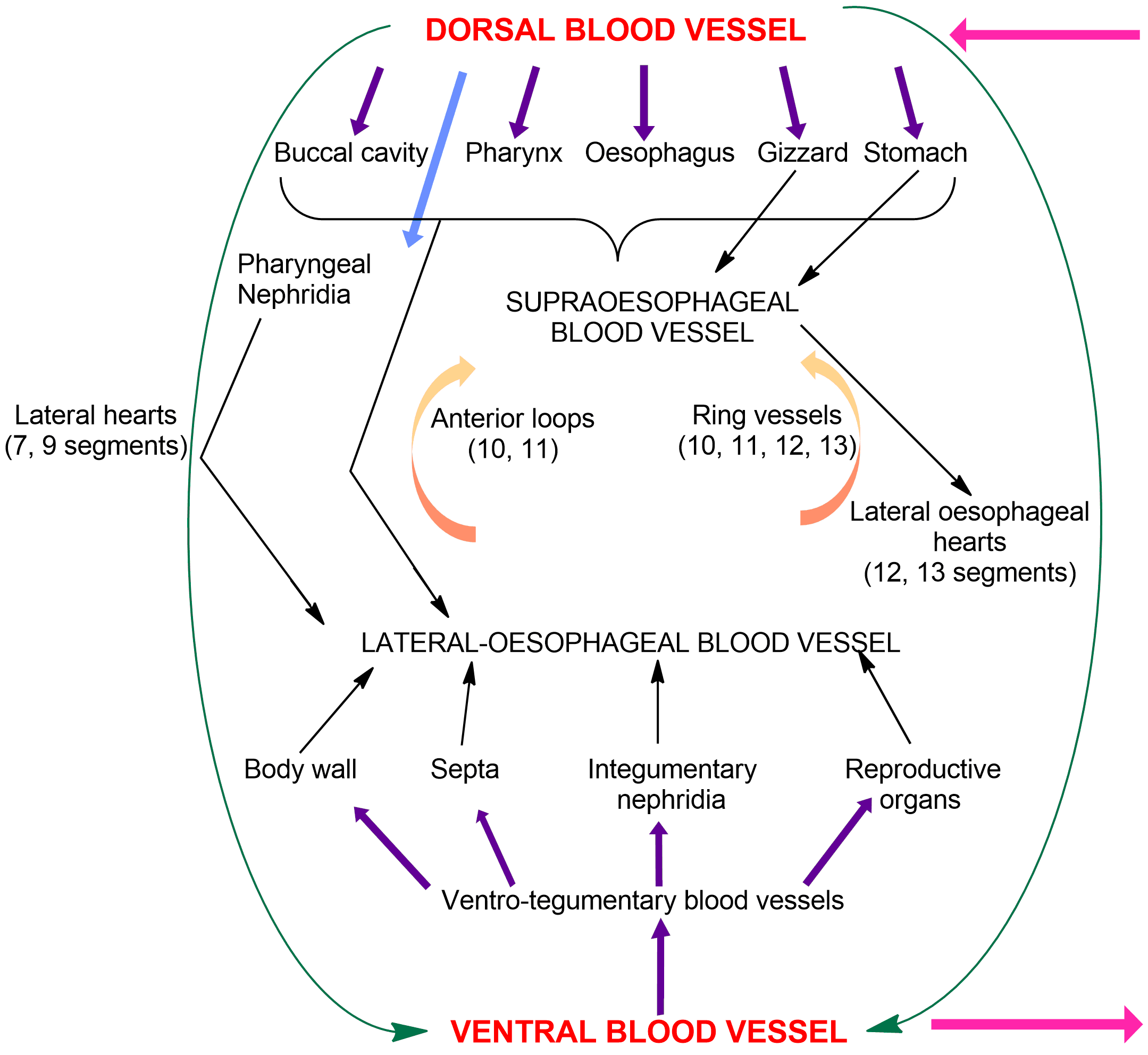
The blood vessels of the circulatory system of earthworm include longitudinal blood vessels, hearts, anterior loops and the ring vessels.
The longitudinal blood vessels: These blood vessels include,
- Dorsal blood vessel- It is the largest vessel in the body extending along the mid dorsal line above the gut. This vessel is muscular, valvular and contractile. In this blood vessel, the blood flows from posterior end to the anterior end. Immediately in front of each septum this vessel has a pair of valves in its lumen. These valves prevent the backward flow of blood. In the first thirteen segments, it acts as a distributing vessel. It sends out the blood collected from the posterior region into the anterior region of the gut and hearts. It extends forward upto third segment where it divides into three branches and distributes to pharyngeal bulb as well as the roof of the buccal chamber. This vessel gives off a pair of stout branches in 3rd, 4th, 5th, 6th and 8th segments. These stout branches take blood to pharynx, pharyngeal nephridia, oesophagus and stomach.
- Ventral blood vessel- The ventral blood vessel, which is the chief distributing blood vessel, lies below the gut along the mid ventral line. It is the longest blood vessel extending upto second segment anteriorly. It is a non-muscular, non-valvular and non-contractile blood vessel which is distributive in nature. In this blood vessel, the blood flows from the anterior end to posterior end, behind the hearts. It gives off a pair of ventro-tegumentary vessels in each segment, which supplies blood to the body wall, septa, integumentary nephridia and reproductive organs present in that segment. The ventro-tegumentary blood vessels lie just in front of the posterior septum of the segment.
- Latero-oesophageal blood vessels- These are a pair of blood vessels which are large and are situated one on each ventro-lateral side of the gut in the first thirteen segments. Two lateral-oesophageal blood vessels are anterior in continuation of sub-neural blood vessel which bifurcates in the 14th segment to formed paired lateral-oesophageal blood vessels. These blood vessels collect blood, through a pair of smaller blood vessels in each segment, from the body wall, septa, integumentary nephridia, pharyngeal nephridia and reproductive organs in the first thirteen segments. They also collect blood from alimentary canal of the first thirteen segments. In these vessels blood flows from anterior to posterior end.
- Supra-oesophageal blood vessel- This vessel is located in the segments from 9th to 13th. It lies on the dorsal side of the stomach. It is a collecting blood vessel and collects blood from the gizzard and the stomach. The supra-oesophageal blood vessel is connected with the lateral oesophageal blood vessels through anterior loops and ring vessels. In this blood vessel, blood flows from anterior end to posterior end. The supra-oesophageal blood vessel at places divides into two and unites again to forma a single vessel.
Hearts: The dorsal blood vessel and the ventral blood vessel are connected by a pair of pulsating hearts, in each of 7th, 9th, 12th and 13th segments. Of these four pairs the anterior two pairs connect only the dorsal blood vessel to ventral blood vessel. Hence they are called as ventral hearts. The posterior two pairs connect both the dorsal blood vessel and the supra-oesophageal blood vessel with the ventral blood vessel. Hence they are called lateral-oesophageal hearts.
These two types of hearts also differ in number and arrangement of their valves. Four pairs of valves are present in each lateral heart, while three pairs of valves are present in each lateral oesophageal heart. Hearts allow the blood to flow into the ventral blood vessel only.
Anterior loops: There are two pairs of loops like vessels, one pair in 10th and 11th segments. These are called anterior loops. They connect the lateral-oesophageal blood vessels with the supra-oesophageal blood vessel. Anterior loops are non-muscular, non-valvular and non-pulsatile. The blood from the lateral-oesophageal blood vessels flows into supra-oesophageal blood vessel through these loops.
Ring vessels: In the segments 10th to 13th, the wall of the stomach shows a series of vessels, connecting the supra-oesophageal blood vessel and lateral-oesophageal blood vessels. They are called ring vessels. They are situated within the muscular coat of the stomach wall. Through these ring vessels, the blood flows from the lateral-oesophageal blood vessels to the supra-oesophageal vessel.
The blood vessels and their arrangement behind first thirteen segments
The blood vascular system behind first thirteen segments consists of longitudinal vessels, commissural vessels and intestinal plexus.
Longitudinal blood vessels: Dorsal blood vessel, ventral blood vessel and sub-neural blood vessel are the three longitudinal blood vessels in this region
- Dorsal blood vessel- It is the collecting blood vessel in this region. It is present along the mid dorsal line above the gut. It collects blood in each segment through dorso-intestinal vessels from the intestine and from the sub-neural blood vessel through the commissural blood vessels. The openings of dorso-intestinal and commissural blood vessels into the dorsal blood vessel are valvular.
- Ventral blood vessel- It is the chief distributing blood vessel in this region. It gives rise to a pair of ventro-tegumentary blood vessels and a single ventro-intestinal blood vessel in each segment. Unlike the ventro-tegumentary blood vessels in the anterior region, they pierce the posterior septum of the segment and extend with their branches into the succeeding segment. The ventro-tegumentary blood vessel travels upwards along the inner surface of the body wall right upto the mid dorsal line and supplies blood to the body wall and integumentary nephridia on its course. Just before it pierces the septum, the ventro-tegumentary gives off the branch called septo-nephridial blood vessel that supplies blood to the septal nephridia. The ventral blood vessel also gives off in each segment, a median ventro-intestinal vessel from its dorsal surface supplying blood to the ventral wall of the intestine
- Sub-neural blood vessel- It extends from 14th segment to the posterior end in the mid-ventral line, beneath the ventral nerve cord. It is non-muscular, non-valvular, collecting vessel. It receives a pair of small branches in each segment collecting blood from the ventral body wall and the ventral nerve cord. The blood in this vessel flows from the anterior to the posterior.
Commissural blood vessels: A pair of commissural blood vessel is present in each segment from 14th onwards. They connect sub-neural blood vessel to the dorsal blood vessel. They collect the blood from the septal nephridia, reproductive organs and the body wall through small branches from 14th till the last segment and send it to the dorsal blood vessel. Each commissural vessel gives off a septo-intestinal branch to the intestine. Thus the commissural blood vessels act as collecting and distributing vessels.
Intestinal plexus: The following are the types of intestinal plexus present in Pheretima.
- Dorso-intestinal blood vessels- Two pairs of dorso-intestinal blood vessels are present in each segment and are located on the dorsal side of the intestine. These vessels collect the blood from the intestinal wall and open into the dorsal blood vessel. The blood in these vessels contains the digested food materials.
- Intestinal blood plexuses- It consists of a network of capillaries in the intestine, one internal and one external. Capillaries of the internal network are situated between the circular muscle layer of the intestine and its internal epithelial lining. The capillaries belonging to the external network lie on the surface of the gut. The internal plexus serves to absorb nutrients from the gut and is connected to the dorsal blood vessel through the dorsal-intestinals. The external plexus receives blood from the ventro-intestinal and septo-intestinal blood vessels and is connected to the internal plexus.

- Write about the importance of circulatory system of Earthworm.
- What is circulation? Describe the process of circulation in Earthworm
- Describe the blood vessels and their arrangement in the first thirteen segments of Earthworm.
- Diffrentiate between the arrangement of blood vessels of the first thirteen segnemts and the segments behind.
- Share with your friends! -
Login to post your comment here...
- or with social Account -