The parasites which complete their life cycle in only one host are called as monogenetic parasites. Also gregarines are the parasites existing in the coelomic epithelial cells, gut and reproductive organs of invertebrate hosts. There are two forms of these sexually reproducing gregarines namely acephaline and cephaline.
In the acephaline forms, the body is not divided into chambers and the anterior end does not bear any organ for the attachment to the host. In cephaline forms, the body is chambered and they also have an organ for attachment to the host. Monocystis is an acephaline gregarine. Monocystis in Greek means mono=single & kystis=bladder. It resides as a parasite in the seminal vesicles of the earthworm
Monocystis species are cosmopolitan in distribution in other words they are found distributed throughout the world.
These are endoparasites in the earth worms. They reside in the coelom and seminal vesicle of the earth worms.
The adult and feeding stage of this parasite is called as trophozoite. It develops within the sperm morula. Sperm morula is a group of developing sperms in the seminal vesicles of earthworm.
External morphology of Monocystis
Shape and size: Trophozoites are small, unicellular and microscopic. The exact size and shape changes according to the developing stage of the trophozoite. Young trophozoites are rounded and oval measuring about 5 µ long. Fully grown trophozoites are elongated, spindle-shaped, flattened and worm like with tapering body ends measuring around 500 µ long and 40 µ broad. This fully grown trophozoite can be viewed with a naked eye.
Pellicle: Pellicle is the external thick and firm structure surrounding the body of the trophozoite. Pellicle may be modified as per the surroundings it may be striated, or ridged or furrowed. The pellicle contains microtubules which are arranged longitudinally.
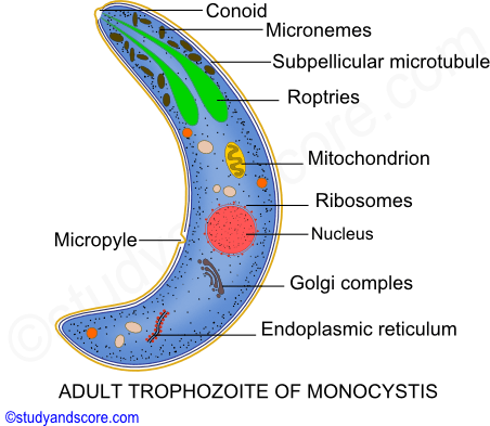
Cytoplasm: The cytoplasm can be differentiated into the outer ectoplasm and the inner endoplasm. The ectoplasm is further divided into outer epicyte, middle sarcocyte and inner myocyte. The inner layer myocyte consists of longitudinal and transverse contractile fibrils called as myonemes. The myonemes assist the trophozoite in metabolic movement. The endoplasm consists of fat globules, granules of special glycogen called Para glycogen. Other organelles in the cytoplasm include mitochondria and Golgi complex.
Nucleus: Nucleus is single with one nucleolus. It may be in elliptical or spherical shape. The nuclear membrane is delicate and bears nuclear pores. Nucleoplasm contains four chromosomes.
Locomotion: Special organs for locomotion are absent in Monocystis. Monocystis moves by wriggling or gliding movement brought about by the rhythmic contraction and relaxation of myonemes. This movement is very much similar to the euglenoid movement of Euglena.
Nutrition: Monocystis absorbs the nourishment from the fluid of the seminal vesicles through the general body surface. So the nutrition of Monocystis is saprozoic type. The reserve food material is stored in the endoplasm in the form of Para glycogen granules.
Respiration: The mode of respiration is by the process of diffusion though its pellicle from the cell contents of the sperm morula. Also the mitochondria synthesize the respiratory enzymes for oxidation reaction for conversion of pyruvic acid to carbon dioxide and water.
Excretion: The resulting waste products of the metabolism like the carbon dioxide and nitrogenous wastes are eliminated out of the body through diffusion into the surrounding fluid of the sperm morula.
Monocystis is monogenetic in other words its lifecycle completes in a single host and it does not require a secondary host like in case of digenetic parasites. The single host of Monocystis is earthworm. All the stages of the life cycle of the Monocystis are haploid except for zygote. The following are stages of the life cycle of the Monocystis.
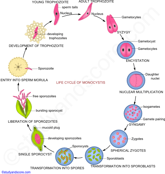
Gamontogamy: This is a sexual reproduction process involving pairing of gamonts (Syzygy), formation of gametes (Gametogony) and fertilization (Syngamy).
Syzygy: The trophozoite in the seminal fluid and coelom of the earthworm wanders about feeding and growing in size and consequently becoming an oval shaped reproductive body called as gamont or gametocyte. These gamonts become shortened and associate with each other in pairs. This pair secretes a protective and resistant cyst wall around themselves. At this stage they are called as gametocyst or gamontocyst. The cyst wall has two layers, the outer called as ectocyst which is rigid and the inner called as endocyst which is thin. The very important point to be noted here is that inside the cyst wall the two gametocytes never fuse. This type of pairing of the gametocytes is called as syzygy
Gametogony: Inside the gametocyst, each gametocyte undergoes nuclear multiplication by mitosis. The nuclei formed after the mitotic division move to the periphery and get surrounded by cytoplasm. These uninucleate cytoplasmic bodies are called as cytomeres. These cytomeres get transformed as gametes and these gametes are called as anisogametes. Anisogametes contain haploid number of chromosomes. All the gametes from one gametocyte are of same sex.
Syngamy: Inside the gametocyst, the gametes mingle and fuse in pairs to produce diploid zygotes. The two gametes uniting come from different gametocytes and are of opposite sex.
Sporogony: The zygote which is initially spherical gets transformed into an oval and unicellular body called as sporoblast. The sporoblast secretes a thick cyst wall call as sporocyst and becomes a spore which is biconical in shape. The spores have a mucoid plug at each end.
Inside the sporocyst, the spores undergo successive nuclear divisions to produce eight daughter nuclei. Each daughter nuclei is surrounded by cytoplasmic mass and is known as sporozoite. All the eight Sporozoites are bundles together in the sporocyst. At this stage the cyst wall ruptures liberating the spores into the cavity of the seminal vesicles of the earthworm.
Transference: The transmission of the Monocystis parasite from one earthworm to other may take place through the following methods. But the exact manner in which the transmission occurs is not known with certainty.
* During copulation
* Death of host
* By birds
* Autotomization
Sporozoites: Sporozoites are minute, spindle shaped protoplasmic bodies. Each sporozoite has a single nucleus, one or more mitochondria, Golgi complex and granules of reserved food. The pellicle covering the body has longitudinally arranged contractile microtubules. A pair of secretory organelles called as roptries are present at the anterior end. The secretion of roptries helps the sporozoite to penetrate through the tissues.
Invasion of the seminal vesicles: The sporozoites move and make their way into the epithelial cells of the gut. Each sporozoite occupies one cell of the gut mucosa. The sporozoites grow in these cells and then escape into the seminal vesicles. After reaching the seminal vesicles each sporozoite enters into the cells of the sperm morula.
Development of trophozoite: Inside the sperm morula the sporozoite feeds and grows into a young trophozoite. These trophozoites have remnants of the degenerating sperms around its body. After the degeneration of these remnants the trophozoite becomes adult and free.

- Share with your friends! -
Login to post your comment here...
- or with social Account -