Protozoans in water are subjected to forces of water resistance like pressure drag and viscous drag. Pressure drag is due to the difference of pressure between two ends of the body. Viscous drag is due to the water molecules attached to the surface of the body.
For protozoans which are small in size, viscous drag is of much importance. These organisms are not streamlined to minimize the pressure drag.
Locomotion is the movement of the animals from place to place. It is performed in search of food, mate, and shelter or to escape from predators etc. it is influenced by external and internal stimuli.
Protozoans are very primitive, single celled animals which show great adaptability in their locomotion. They exhibit slowest locomotion like amoeboid locomotion and also the fastest locomotion like ciliary locomotion.
In protozoans, locomotion is brought about by
They are also known as false feet. These are the temporary outgrowths of the cell. They are formed on the surface of the body by the movement of the cytoplasm.
Polypodia- Several pseudopodia formed on the surface of the body.
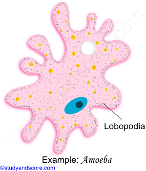
Eg: Amoeba proteus
Monopodia- Only single pseudopodia is formed on the surface of the body.
Eg: Entamoeba histolytica
Lobopodia: These are lobe like and blunt structures with broad and rounded ends. These structures composed of endoplasm and ectoplasm. Lobopodia move by pressure flow mechanism.
Eg: Amoeba proteus, Entamoeba histolytica
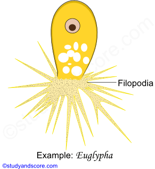
Filopodia: These are slender filamentous pseudopodia tapering from base to tip. Sometimes these may be branched out but they are not fused to form a network. They are composed of only ectoplasm.
Eg: Euglypha, Lecithium
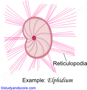
Reticulopodia: They are also known as rhizopodia or myxopodia. They are filamentous, profusely interconnected and branched. They form a network. The primary function of these pseudopodia in ingestion of food and the secondary function is locomotion. They exhibit two way flow of the cytoplasm. They are commonly found in foraminifers.
Eg: Elphidium, Globigerina
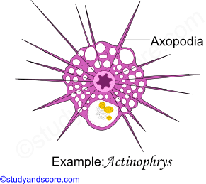
Axopodia: These are fine needle like, straight pseudopodia radiating from the surface of the body. Each Axopodia contain a central axial rod which is covered by granular and adhesive cytoplasm. The main function of these axopodia is food collection. Axopodia also exhibit two-way flow of cytoplasm. Axopodia are mainly found in Heliozoans and radiolarians.
Eg: Actinosphaerium, Actinophrys, Collozoum
Many protozoans have contractile structures in the pellicle or ectoplasm called as myonemes. These may be in the form of,
* Ridges or grooves (Eg: Euglena)
* Contractile myofibrils (Eg: Larger ciliates)
* Microtubules (Eg: Trypanosoma)
Flagella are the locomotory organelles of flagellate mastigophoran protozoans. They are mostly thread like projection on the cell surface. A typical flagellum consists of an elongated, stiff axial fiber called as axial filament or axoneme enclosed by an outer sheath. The axoneme arises from basal granule called as blepharoplast or kinetosome which is further derived from Centrioles. Blepharoplast lies below the cell surface in the ectoplasm. The region around blepharoplast is called microtubular organizing center that controls the assembly of microtubules.
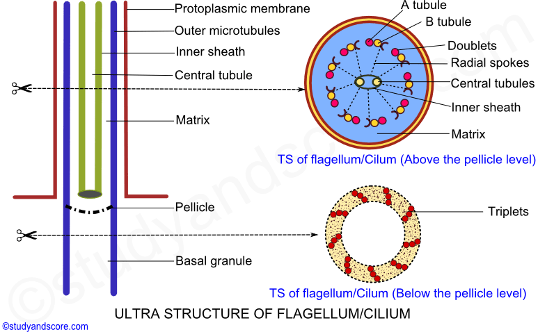
When the axial filament is viewed under an electron microscope 9 + 2 arrangement can be observed. The 2 central longitudinal fibers are enclosed by membranous inner sheath. The 2 central longitudinal fibers are surrounded by 9 longitudinal peripheral doublets (each with microtubules A and B) which form a cylinder between the inner and the outer sheath. Each peripheral paired fiber is connected to the internal membranous sheath by radial spokes.
Each peripheral doublet also has pairs of arms directed towards neighboring doublet. These arms are made of the protein called as dynein. The arms create the sliding force. The peripheral doublets are surrounded by an outer membranous sheath called as protoplasmic sheath, which is an extension of the plasma membrane. Some flagella also bear lateral appendages called as flimmers or mastigonemes along the length of the axoneme above the level of the pellicle.
Number and arrangement of flagella vary in Mastigophora from one to eight or more. Free living species usually have one to eight flagella whereas the parasitic forms may have one to many flagella. Flagella are classified based on the arrangement of lateral appendages and the nature of the axial filament.
Stichonematic: Only one row of lateral appendages occurs on the axoneme up to tip.
Eg: Euglena, Astasia
Pantonematic: Two or more rows of lateral appendages occur on the axoneme
Eg: Peranema, Monas
Acronematic: Lateral appendages are absent and axoneme ends as a terminal ‘naked’ axial filament
Eg: Chlamydomonas, Polytoma
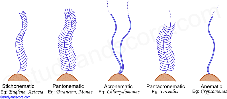
Pantacronematic: Flagellum is provided with two or more rows of lateral appendages and the axoneme ends in a terminal naked axial filament.
Eg: Urceolus
Anematic: In some cases the flagella is simple without any lateral appendages and a terminal naked filament.
Eg: Chilomonas, Cryptomonas
Cilia are short hair like structures present all over the surface of the body. They may be also confined to specific regions of the ciliate protozoan. Cilia help in locomotion as well as in food collection.
Cilia greatly resemble the flagella in the basic structure. The major difference between the flagella and the cilia is that cilia are smaller compared to the flagella. Cilia arise from the kinetosome. Cilia consist of an axial filament called as axoneme surrounded by the protoplasmic outer sheath.
Electron microscopic studies of axoneme reveal 9 + 2 organization of the peripheral doublet fibrils and central singlet fibrils. The details of the 9 + 2 organization and the presence of the dynein arms are similar to that of the flagellum. All these fibrils are embedded in a matrix. The central fibrils are enclosed within a delicate sheath.
The infraciliary system is located just beneath the pellicle. It consists of kinetosomes at the bases of cilia, kinetodesmos or kinetodesmal fibrils that are connected to the kinetosomes and running along the right side of each row of kinetosomes as cord of fibers known as kinetodesmata. A longitudinal row of kinetosomes, kinetodesmal fibrils and their kinetodesmata form a unit called kinety. All the kineties together form an infraciliary system that lies in the ectoplasm. The infraciliary system is connected to the motorium, a neuromotor center neat the cytopharynx and forms the neuromotor system. This neuromotor system controls and coordinates the movement of cilia.
* In some primitive forms like holotrichs (Eg: Paramecium) cilia are present all over the body
* In some forms like peritrichs (Eg: Vorticella) cilia are present only in the peristomial region
* In Suctorians (Eg: Acineta) cilia are present in only in the young ones which are later replaced by sucking tentacles in the adults
| Flagella | Cilia |
|---|---|
| They may be one to four in number. Mor than four flagella are present in mastigophoran parasites | Generally cilia are more in number compared to flagella. Cilia vary from 3,000 to 14,000 in number |
| A flagellum is about 150 microns in length | A cilium is about 5 to 10 microns in length |
| Flagella are commonly found at one end of the cell | Cilia occur either all over the body surface or at specific regions of the cell |
| Flagella produce undular movement | Cilia produce pendular movement |
| Flagella help in locomotion only | Cilia help both in feeding and in locomotion |
| Flagella do not form compound organelles | Cilia may form undulation membranes and other compound ciliary organelles |
* Cilia in compound ciliary organelles do not fuse, but their basal granules are sufficiently close to introduce a sort of coupling.
* A group of cilia that forms a bundle is called as cirrus. An undulating membrane is a row of adhering cilia forming long sheet.
* The smaller rows of adhering cilia form the membranelles

- Share with your friends! -
Login to post your comment here...
- or with social Account -