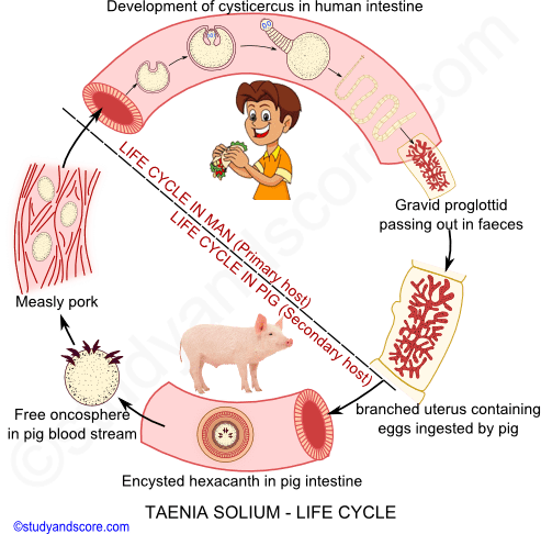
The life-history of Taenia solium is complicated and digenetic. It is completed in two hosts. The primary host is man and the secondary host is pig. There is no free larval stage in the life cycle of Taenia.
Self-fertilization takes place in Taenia. Fertilization is preceded by copulation. During copulation, the cirrus of the segment is inserted into the vagina of the same segment. The sperms received are stored in the seminal receptacle. The eggs are fertilized in the oviduct and get surrounded with yolk and egg-shell in the ootype.
The capsulated egg enters the uterus and is collected there. The uterus enlarges in size, gets branched and occupies the whole space. The eggs are very small in size measuring about 40 microns in diameter. These contain a large amount of yolk and each is surrounded by an egg-shell or egg-capsule. The embryonic development begins in the uterus itself.
The division in the eggs starts, while still inside the uterus. The first cleavage is unequal so that a large megamere and a small embryonic cell are formed. The embryonic cell undergone repeated divisions and a solid ball of cells, the morula is formed. The divisions are unequal so the morula consists of a few larger cells, the macromeres or mesomeres forming an outer or peripheral layer and inner mass of small cells or micromeres.
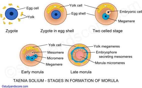
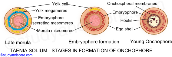
The mesomeres surrounding the embryo form an inner embryonic membrane or embryophore. The megamere also divides to form outer embryonic membrane outside the embryophore. The megamere provides nourishment to the growing embryo.
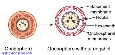
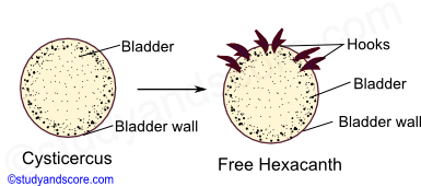
The micromeres develop into a hexacanth or oncosphere. The oncoblasts of the embryo secretes three pairs of chitinous hooks. This embryo with six hooks surrounded by two embryonic membranes is called as hexacanth larva or oncosphere. 30,000 to 40,000 oncospheres are present in each gravid proglottid and such a gravid proglottid with such huge numbers of oncospheres get detached from the main parasite body by the process of apolysis and pass out through the faeces.
Infection to secondary host takes place through the oncosphere stage. The gravid proglottid containing oncospheres are present in the infected human faeces. When a pig feeds on such faecal matter, the gravid proglottids enter the stomach of the pig and get released.
The shell and embryophore are dissolved in the duodenum of the pig. Now the larvae are set free to pass into the intestine and get attached to the intestinal mucous layer with the help of the hooks. These larvae later penetrate the intestinal wall with the help of the substances produced by the penetration glands.
Later, the hexacanth larva enters the hepatic portal vein, enters the heart and gets settled in the voluntary muscles of tongue, elbows, limbs, neck. Sometimes it may also settle in the organs like lungs, eyes, kidneys and brain.
The hexacanth loses its hooks in the voluntary muscles of the pig and develops into a pre-adult stage called as cysticercus or metacestode phase. The early cysticercus absorbs nutritive substance from host tissues and grows in size. A cavity appears in the center of its cell mass due to the degeneration of the mesenchymal cells.
This cavity gets filled with fluid containing plasma of the pig. At this stage the embryo is surrounded by two layers namely the outer cuticle and the inner mesenchymal layer or germinal layer.
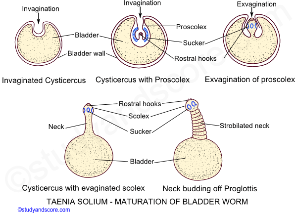
The wall of the embryo gets thickened and invaginates into tits cavity in the form of a knob. Internally this knob develops four suckers, a small rostellum and hooks at its base. It is called as proscolex. The embryo is now called mature cysticercus or hydatid larva or bladder worm. It has ellipsoid milky white bladder surrounded by fibrous capsule. These cysts are called cysticercus cellulosae. The pork with these cysticercus cellulosae is called measly pork
Further development of the bladder worm takes place only inside the definitive host. Infection of man occurs when inadequately cooked pork infected with bladder worms is eaten. The cysticercus becomes active in the intestine. The scolex takes a firm hold of intestinal wall of the host. The bladder is thrown off and the neck starts budding off segments an adult tapeworm is formed.

- Share with your friends! -
Login to post your comment here...
- or with social Account -