The life history of Ascaris includes many stages namely, Copulation, Fertilization, Formation of Zygote, Cleavage and early development, infection to new host, later development in the new host and finally migration. All the stages are described hereunder:
The copulation takes place in the small intestine of humans in which the adult roundworms reside. The male worm orients its body at right angles to that of the female worm so that its cloacal aperture opposes the vulva of the female. The penial spicules of the male worm help in opening the vulva of the female worm. After the opening of the vulva, the sperms are transferred into the vagina from where the sperms are passed on to the proximal end of the uteri.
After the sperms reach the proximal end of the uteri, the process of fertilization begins. After fertilization, the ova undergo second maturation division.
The eggs which are not fertilized contain glycogen and fat. After fertilization, the glycogen globules migrate to the surface and form a fertilization membrane which later hardens to form thick, clear and chitinous shell. Now the fat globules form a thin lipoid layer below the shell.
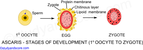
As the zygote moves down the uterus, the uterine wall secretes a thick hard, yellow or brown aluminous coat with a typical wavy texture. The fertilized eggs at this stage are elliptical in shape. Under appropriate conditions of temperature, moisture and oxygen the fertilized eggs undergo cleavage to develop into infective stage.
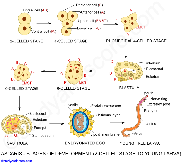
The cleavage in roundworms is determinate and spiral type. The first division results in two cells namely dorsal (AB) and ventral (P1). Next the dorsal cell divides into anterior A and posterior B, whereas the ventral cell divides into upper EMST and the lower P2. The four-celled embryo thus formed is initially in T-shape but soon it becomes rhomboidal in shape as the P2 comes to lie posterior to EMST.
In the next cleavage,
* A and B further divide in to right and left cells,
* EMST divides into E and MST,
* P2 divides into P3 and C
Further,
* E divides into E1 and E2
* P3 divides into p4 and D
Finally,
* P4 divides into G1 and G2
|
The fate of various cells is pre-determined and fixed as follows, * The descendants of A and B give rise to ectoderm * MST gives rise to mesoderm and ectoderm of foregut * E1 and E2 gives rise to endoderm * G1 and G2 form germ cell primordium * C and D together give rise to ectoderm and mesoderm |
After all the divisions, embryo attains 16 cells and forms a hollow ball like structure called as blastula. The cavity of blastula is called as blastocoel. Blastula undergoes invagination process also called as gastrulation to form gastrula. The gastrula grows in size and gets transformed into an active juvenile with alimentary canal, nerve ring and a larval excretory system. This juvenile closely resembles the nematode genus Rhabditis, found in soil and human faeces. Thus this juvenile is also known as rhabditiform larva of first stage. This stage is not infective. In a week time this larva undergoes moulting within the egg shell to form second stage larva which is infective.
As there is no intermediate host in the life cycle of Ascaris, humans acquire infection by
a) directly ingesting Ascaris eggs, with second stage larva,
b) contaminated food or water
After ingestion of the eggs, due to the action of the digestive juices of the small intestine, the egg shell of the larva is dissolved and the larva hatch out.
Just hatched out juvenile measures about 0.25 to 0.3 mm in length and 13 to 15µ in diameter. This juvenile performs active thrashing movements to bore through the intestinal epithelium and begins to migrate to other parts of the host body.
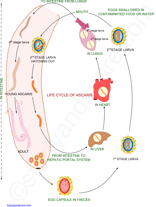
The larva migrates the hepatic portal circulation and then to the liver and then to heart through post-caval vein. From the heart it migrates to the lungs via pulmonary artery. In the lungs it remains for few days and increases in size. It ruptures the blood capillaries and bores its way to alveoli. Here this second larval stage undergoes moulting to become third stage larva. This third stage larva further undergoes another moulting to transform into fourth stage larva which is about 2-3 mm in length.
This fourth stage larva migrates from the alveoli to the pharynx through trachea. From the pharynx this larva reenters the gut when coughed and swallowed back. This fourth stage larva undergoes final moulting to become adult. These adult worms attain sexual maturity within 8-10 week time.
Some larvae do not follow the above said usual pattern of migration but reach the brain or spinal cord. The larva cannot survive in these organs and thus a calcareous cyst is formed around it.

1. Write about cleavage and early development in the life history of Ascaris.
2. Draw a neat labelled disgram of life cycle of Ascaris.
- Share with your friends! -
Login to post your comment here...
- or with social Account -