Fasciola hepatica is hermaphrodite. The female and male genital ducts open into common chamber called genital atrium. The gonads in these liver flukes are well developed. The genital atrium is situated in the anterior part of the body and it open out through common genital aperture or gonopore. The gonopore is located just in front of the acetabulum.
The following are the organs of the male reproductive system,
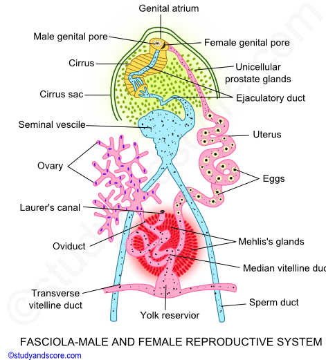
The following are the parts of the female reproductive system,
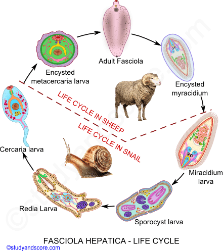
The life cycle of Fasciola hepatica is complex and it is completed in two different hosts as it is a digenetic parasite. The primary host is sheep in which the adult liver flukes live. Whereas the intermediate or secondary host is a snail in which all the larval stages are developed. The following are the stages in the life cycle of Fasciola hepatica.
Though liver flukes are hermaphrodite, they undergo cross fertilization preceded by the process of copulation. Self-fertilization occurs very rarely. The copulation of Fasciola hepatica takes place in bile ducts of the host. The two flukes in copulation bring their general pores opposite to each other. The cirrus of one fluke everts out through the gonopore and penetrates through the Laurer’s canal of the other fluke and injects the spermatozoa. The secretions of the Mehlis’s glands and prostate glands help in fertilization.
The fertilization in these liver flukes is internal type. In cross fertilization, the sperms received in the Laurer’s canal during the copulation, enter the distal end of the oviduct. And in the oviduct the fertilization takes place. And in self-fertilization, the sperms enter the uterus of the same fluke through female genital aperture and pass down to fertilize the eggs.
Each of the fertilized eggs is surrounded by the yolk cells. These yolk cells provide nourishment and shell material to the developing eggs. The shell globules of the yolk cells contain protein and a phenol.
All the shelled eggs are called as capsules. The shell of the capsule is yellow or brown in color and oval in shape. The capsules are also provided with a lid called as operculum. Beneath the operculum a viscous and granular cushion is present. About 200 flukes are present in one sheep and each of these flukes produces around 500,000 eggs and a single infected sheep may disperse 100 million fertile eggs. Such a vast capacity of egg production is necessary as the life of these flukes is complicated and there are very less survival chances.
While the eggs are still in the uterus, the process of cleavage starts. The cleavage is holoblastic and unequal type. First the zygote divides to form two unequal cells; the larger called the somatic cell and the smaller called propagatory cell. Somatic cell divides further to form larval ectoderm and tissues. Propagatory cell divided further to produce two daughter cells. One of these daughter cells produces the larval body and the other daughter cell undergoes further divisions to form mass of germ cells. These germ cells cluster in the posterior part of the larval body.
These embryos get capsulated and cannot develop further in the fluke’s uterus. Numerous capsules leave the fluke’s body through the gonopore into the host’s intestine and are finally ejected out through its faeces. The further development of these capsules takes place when these come in contact with the water which is slightly acidic.
When the conditions turn favorable, the encapsulated embryos get differentiated into miracidium larva. It is the first larval stage in the life cycle of liver fluke. These larvae are hatched out with the help of hatching enzyme. This enzyme dissolves the cementing material of the operculum.
External structure of miracidium larva: Miracidium larva is small, oval, elongated and richly ciliated. The anterior end produces mobile, non-ciliated apical papilla. This larva is multicellular. Its body is covered with ciliated epidermal plates which are total 21 in number arranged in five rows or tiers. The number of plates in each tire is fixed.
Under these epidermal plates, a fine layer of sub-epidermal musculature is present. Thus, musculature consists of outer circular and inner longitudinal fibers. Under this musculature another layer called sub-epithelium is present. Epidermal plates, sub-epidermal musculature and sub-epithelium together form the body wall of miracidium.
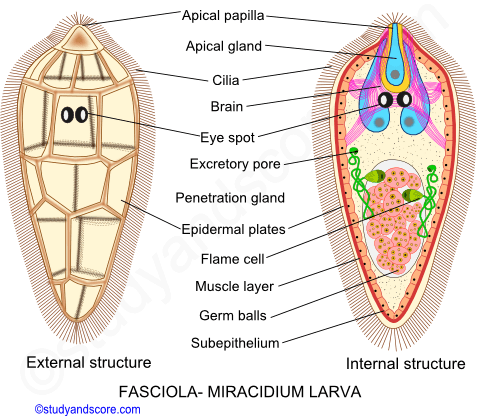
Internal structure of miracidium larva: Inside the body of miracidium, numerous glands, nervous tissue and protonephridia and germ cells are present. A sac-like multinucleated mass of granular protoplasm is attached to the center of apical papilla. A large brain with several associated nerve fibers lies dorsally below the epidermal cell of the second tier. Above the brain, an “X” shaped larval eye with two crescentic pigmented cells called as eye spots are present. The concavities of these eyes face each other. These concavities contain refractile material serving as lens. A pair of long tubular protonephridia or flame cells opens to the exterior through excretory pores. The germ cells lie in groups called as germ balls in the rear part of the body of miracidium larva.
Miracidium larva does not feed but swims about desperately are tries to penetrate any object it may come across. But it can succeeds only if it comes in contact with the specific intermediate host, snail. All the larvae which do not come in contact with the suitable host would die within 24 hours. After finding the suitable host, the larva attaches itself with the apical papilla and performs boring movements. This movement along with the flesh-dissolving secretions of the larva, it created a minute bore in the host tissue. Through this minute the larva squeezes itself into the host body. After entering the body of the host it sheds of its ciliated epidermis and then enters into the digestive gland of the sheep to undergo further changes and finally develops into next larval stage called as sporocyst larva.
Sporocyst larva is an elongated structure which retains the body layers of the previous larval stage except the ciliated epidermis. This ciliated epidermis is replaced by a thin cuticle. The glands, brain, eye spots and apical papilla which are present in the miracidium larva get degenerated in this stage.
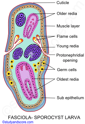
The protonephridia of both the sides divides into two flame cells which finally open out through a common excretory pore. This larva moves about in the host tissue absorbing nutrition. The germ balls present in the sporocyst further develop in to the next larva form called as Redia.
The germ balls present in the sporocyst larva develop into redia larva. The redia larvae come out of the sporocyst by rupturing its body wall. Redia larvae are elongated and cylindrical structures. They have mouth at the anterior end, also a ring of collar cells along with the birth pore are present at the anterior end. The body wall of this larva consists of cuticle, circular, longitudinal muscles and sub-epithelium. The mouth leads into pharynx which further leads into intestine lines by single later of cells. Numerous pharyngeal glands open into pharynx. Protonephridia are highly branched and all the flame cells open out through a common excretory pore. The body of this larva is packed with germ balls and mesenchyme cells.
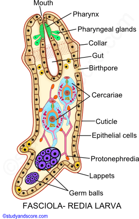
Redia larva moves and feed through the host tissue. The movement of this larva is brought about by the muscular contractions assisted by collar and lappets. This moving redia larva prefers to move into the digestive glands. During summer, when sufficient nourishment is available, the germ balls of this larva give rise to second generation of redia larvae. And during winter season, the germ balls of rediae of second generation develop into next larval stage called as Cercaria larva.
Each of the Redia larvae of the second generation gives rise to around 20 cercaria larva. They leave the body of redia from the birth pore present at the anterior end and then enter the digestive glands of the snail. Cercaria larva is oval in structure with a long and simple tail. The tail helps in swimming movement of this larva. The body layers are much similar to the previous two larval forms. Cuticle has backwardly directed spines. Body wall consists of numerous cystogenous gland cells which secrete cyst for the next larval stage.
Mouth leads into pharynx, followed by oesophagus and intestine. At the lateral zones, numerous flame cells are present, these open into a pair of excretory tubules which further unite to form an excretory vesicle. Groups of germ cells lie in the body which represents the rudiments of adult genital system.
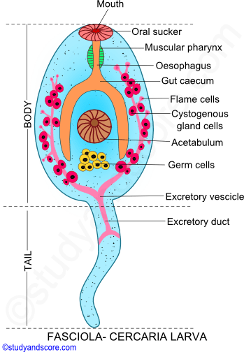
A matured cercaria larva makes its way out of the host tissue, migrates to the pulmonary sac and then from there it escapes in to the surrounding water. After the entry of the miracidium larva into the body of the snail, it takes almost about 35-65 days to exit as cercaria larva.
The cercaria larva swims about in the water and gets attached to grass blades or other aquatic plants. After some time this larva loses its tail and gets encysted to form Metacercaria larva.
The thick, hard and whitish cyst of the metacercaria larva is secreted by the cystogenous gland cells. After the secretion of the cyst these gland cells degenerate. This encysted larva must be ingested by the primary host again. If an uncysted larva is ingested by the sheep they would be digested by the acidic action of the sheep’s gastric juice.
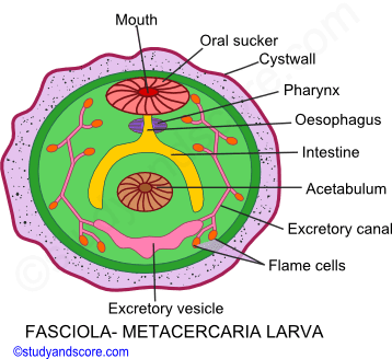
Metacercaria larva has rounded structure. This larva is a juvenile fluke also called as marita. This larval form has large number of flame cells. The cyst of this larva also protects it from desiccation.

- Share with your friends! -
Login to post your comment here...
- or with social Account -