Leishmania sps are the cause of various diseases in man, cattle, dogs, sheep, horse etc. collectively known as leishmaniasis. It is a pathogenic zooflagellate and is closely related to Trypanosoma. These pathogens are carried by blood sucking sand flies of the genus Phlebotomus. Leishmania species are intracellular parasites in the white blood cells, liver cells and spleen cells.
Distribution
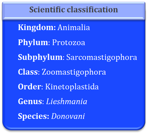
Leishmania donovani is known to infect man in India, China, South America, parts of Africa and Mediterranean countries. This genus was created by Ronald Ross in 1903. The species was simultaneously reported by Leishman in London and Donovan from Madras (India), hence the Leishmania donovani.
Habit and habitat
It lives as an intracellular parasite in the leucocytes or the organs of the reticulo-endothelial system like bone marrow, spleen, liver and lymphatic glands etc. This parasite causes disease called as kala azar fever in humans with the symptoms like enlargement of spleen and reduction in the number of white blood cells. It is transmitted through the bite of Sand flies.
Morphology
Size and shape: The genus Leishmania occurs in two forms namely leishmanial and leptomonad. These two forms alternate between a vertebrate and an invertebrate host. These two forms are recognized on the basis of the position of the kinetoplast and blepharoplast. Also the course taken by the flagellum is important in identification of the leishmanial form.
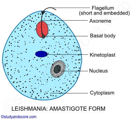
Leishmanial form- This form is also known as the amastigote form. The size ranges from 2 µ to 4 µ in diameter. This form occurs intracellularly in the blood cells and the organs of the reticulo-endothelial system like liver, spleen, bone marrow of man. This form is microscopic, rounded or oval with a central nucleus, blepharoplast and kinetoplast. This form does not have a free flagellum.
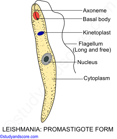
Leptomonad form- This form is also known as the promastigotes form. Its size ranges from 15 – 20 µ in length and 1-2 µ in width. This form occurs in the midgut of the invertebrate host or sand fly. This form is elongated, slender and spindle shaped with a large centrally placed nucleus, blepharoplast and kinetoplast. These forms possess a free flagellum.
Cell membrane: The whole body is covered externally by a very thin, delicate, elastic and firm covering called as pellicle. This pellicle gives a definite shape to the body.
Flagellum: Leishmania is uniflagellate that is it has only single flagellum. In the leptomonad form the flagellum is long and free. This flagellum arises from a minute basal body or blepharoplast situated near the anterior end. Near the blepharoplast a disc-shaped parabasal body called as kinetoplast is present. In the leishmanial form, there is no free flagellum rather the flagellum is reduced and embedded in the cytoplasm. Embedded
Cytoplasm: Homogenous and colorless cytoplasm is present in this parasite. The cytoplasm is not differentiated into ectoplasm and endoplasm. The cytoplasm is marked by longitudinal striations or microtubules. Many other structures like Golgi body, mitochondrion, vacuole, nucleus, blepharoplast and kinetoplast are present in the cytoplasm.
Nucleus: The nucleus of Leishmania is spherical and lies in the middle of the body. It has a central distinct nucleolus. The nucleus is covered by a double unit membrane with pores of size 1 µ in diameter.
Metabolism
Leishmania do not have a mouth or a cytostome and so the nourishment is obtained saprozoically through the general body surface from the host cells. The exchange of the gases and elimination of the waste products takes place by the process of diffusion. Only asexual reproduction takes place in Leishmania through longitudinal binary fission.
Leishmania is a digenetic parasite which requires two hosts to complete its life cycle. These two parasites are named as primary and secondary hosts. The primary host is the principal host which is a vertebrate or man. In the primary host the parasite feeds and multiplies itself asexually. On the other hand the secondary host is the intermediate host or vector which is usually an invertebrate or a blood sucking insect. In the case of Leishmania it is sand-fly (Tse-tse fly). This sand fly belongs to the genus Phlebotomus.
In the life cycle of Leishmania we can also find a reservoir host which can be mammals like dog, jackals and ground squirrels. In the reservoir host the parasite does not undergo any change rather it simply waits to find its principal host through sand fly.
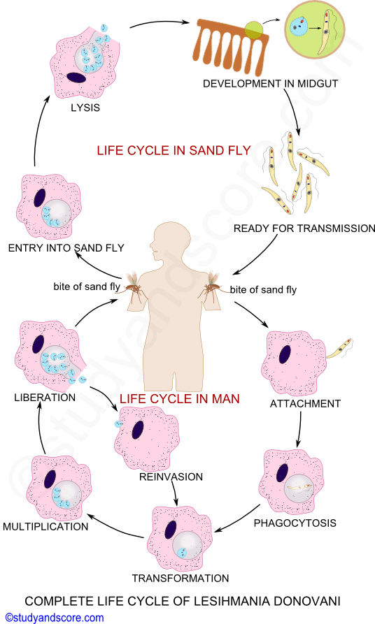
Life cycle in Man
In India, Leishmania donovani is transmitted to man by the infected sand fly (Phlebotomus argentipes). The sand fly injects the infective stages called as promastigotes or leptomonad forms from their proboscis into the human blood. Some of them which enter the blood circulation are phagocytized by the macrophages and other mononuclear phagocytic cell. Some of promastigotes enter the cells of reticulo-endothelial system (consists of the organs like spleen, bone marrow, liver and lymph nodes). Now these promastigotes forms get transformed into amastigote or leishmanial form. These amastigotes get settled in the organs of the reticulo-endothelial system and undergo slow multiplication by binary fission and because of this the respective organs become significantly enlarged.
When the number of parasites reached 50 to 200 or more the macrophages get ruptured to liberate the parasites. Now the liberated parasites are taken up by new host cells and the cycle of multiplication repeats consequently the reticulo-endothelial system progressively becomes infected.
Some of the free amastigotes become phagocytized by neutrophils and monocytes. These heavily parasitized cells wander through the general blood circulation leading to the general infection.
Life cycle in Sand fly
When a sand fly sucks on the blood of an infected person, it obtains free amastigotes and parasitized neutrophils and monocytes along their blood meal. The amastigote forms develop in the midgut of the sand fly. They get transformed into elongated, free flagellated promastigotes forms again. These promastigotes forms multiply by longitudinal binary fission. In about a weeks’ time the number of parasites become enormous thus spread to other organs of the sand fly like pharynx and buccal cavity. The important point to be noted in vector is that the salivary glands are not infected. The transmission to the host takes place when a heavily infested sand fly bites a new host.
Occurrence: Dumdum fever is the serious oriental disease of man caused by Leishmania donovani and transmitted by Phlebotomus argentipes. It is also known as black fever or Kala-azar. The word kala-azar is derived from two Indian words Kala meaning black and azar meaning sickness. This fever is prevalent in India, China, Mediterranean countries and some parts of Arica and South America.
Symptoms and pathogenesis: The incubation o period of dumdum fever is long ranging from 3 to 6 months. Also the symptoms of the fever may appear even after 2 years. The very early symptoms of dumdum fever include swelling, high fever, enlargement of organs or reticulo-endothelial system like spleen and liver. These symptoms are followed by general weakness, anemia due to reduction in the number of blood cells and darkening of the skin.
In the advanced stages the skin of the infected person becomes completely dry, rough and dark with lot of pigmentation, hair becomes brittle and fall out. If the patient does not receive proper treatment it can lead to his death in 2 years. Generally death is due to the secondary infection by bacteria or virus as the patient becomes immune-compromised in other words, the patient would not be able to resist the infections caused by bacteria or virus or any other microbes.
Diagnosis: Microbial examination of the blood film or the biopsy material taken from the spleen or bone marrow of the patient are the main diagnostic samples used for the identification of the infection in a person. Amastigote forms of Leishmania donovani are tested in these samples. Also the examination of the white blood cells shows decrease in the number of neutrophils and increase in number of lymphocytes and monocytes. The number of red blood cells is also found to be decreased.
Treatment: Generally two groups of drugs are used for the treatment of dumdum fever. These include pentavalent antimony compounds like sodium-antimony tartrate and sodium-antimony glutamate, urea stibamine, aminostiburea along with pentamidine isethionate.
Prevention: The following are the prophylactic measured to be taken to prevent the disease
* Eradication of the insect vector is of prime importance to keep the disease under control. Low trees and bushes should be cleared out in the endemic areas. Periodic fumigation and spraying of the insecticides is also advisable to prevent the disease.
* Reservoir hosts like dogs, jackals, squirrels should be kept at bay.
* Proper treatment campaign is a good control measure in India.
* To avoiding the bites of sand fly insect repellants, mosquito nets can be used. Also avoiding to sleep on the floor can be beneficial.
| Species | Primary host | Secondary host | Location in host | Transmission | Disease name | Symptoms |
|---|---|---|---|---|---|---|
| L. donovani | Cats, Man and dogs | Sand fly | Intracellular in WBC, liver & spleen cells, Bone marrow & lymph glands | Bite of sand fly | Kala-azar or dumdum fever or black fever | Fever, Anemia, enlargement of spleen and liver, darkening of the skin |
| L. infantum | Children | Sand fly | Intracellular | Bite of sand fly | Infantile kala-azar | Enlargement of spleen |
| L. chagasi | Man | Sand fly | Intracellular | Bite of sand fly | South American Kala-azar | Similar to Kala-azar |
| L. canis | Dogs | Sand fly | Intracellular | Bite of sand fly | Visceral leishmaniasis | Similar to Kala-azar |
| L. tropica | Man and dogs | Sand fly | Intracellular in skin | Bite of sand fly | Oriental sores | Lesions on the skin |
| L. braseliensis | Cats, Man and dogs | Sand fly | Intracellular in mucous membrane of nose & throat | Bite of sand fly | Naso-pharyngeal leishmaniasis | Infection in the nasopharyngeal region |

- Share with your friends! -
Login to post your comment here...
- or with social Account -