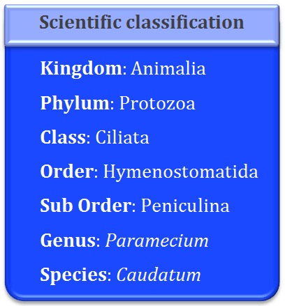
It is worldwide in distribution. It is found in fresh water ponds, pools, streams, lakes, rivers etc. It is found in abundance in stagnant water bodies containing dead and decaying organic matter. It is commonly called as slipper animalcule due to its shape which is like slipper.
Size: Paramecium is a unicellular microscopic protozoan. The largest species of this genus is Paramecium caudatum and it measures about 170-290 µ. It is visible to naked eye as whitish or grayish spot. The greatest diameter of the cylindrical body is about two-thirds of its entire length.
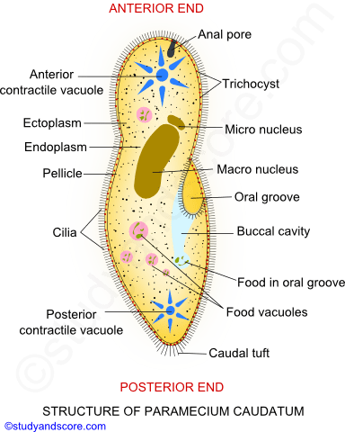
Shape: It is elongated, slipper-shaped animal and is commonly called as slipper animalcule due to its shape. The anterior end is rounded and the posterior end is cone-shaped. Ventral or oral surface is flat and the dorsal or aboral surface is convex.
Pellicle: The body of this protozoan is covered externally by a colorless, thin, firm and elastic cuticular membrane called as pellicle. It is also known as periplast. The firm pellicle provides definite and constant shape to this protozoan and also allows bending movements. Pellicle contains large number of small polygonal or hexagonal depressions or ciliary fields. The marginal hexagonal depressions are slightly raised up as ridges. Trichocysts open in these ridges. A cilium comes out through the center of each depression.
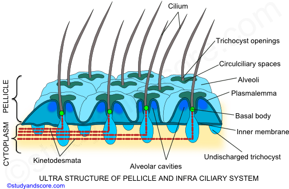
According to the electron microscopic studies, pellicle is composed of three layers. The outermost layer is called as cell membrane. It covers the cilia also. Second and third layers of the pellicle are called as outer alveolar and inner alveolar membranes. The two alveolar membranes enclose a series of alveolar spaces or cavities in between them.
Oral groove: The ventral surface of the body of this protozoan bears a prominent, oblique and shallow depression. This depression is known as oral groove. It originates from the middle of the body and extends to the left side of anterior end. At the posterior side the oral groove leads into a deeper conical vestibule which in turn communicates with buccal cavity.
Cilia: Cilia are short hair like structures present all over the surface of the body. They may be also confined to specific regions of the ciliate protozoan. Cilia help in locomotion as well as in food collection. In Paramecium, the entire body surface is covered by numerous, tiny hair. These cilia are arranged in longitudinal rows throughout the body. The length of the cilia is uniform but a few longer cilia are present at the posterior end. These cilia at the posterior end form the caudal tuft in P. caudatum.
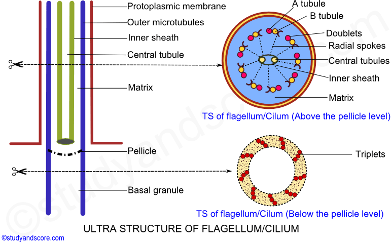
Cilia greatly resemble the flagella in the basic structure. The major difference between the flagella and the cilia is that cilia are smaller compared to the flagella. Cilia arise from the kinetosome. Cilia consist of an axial filament called as axoneme surrounded by the protoplasmic outer sheath.
Electron microscopic studies of axoneme reveal 9 + 2 organization of the peripheral doublet fibrils and central singlet fibrils. The details of the 9 + 2 organization and the presence of the dynein arms are similar to that of the flagellum. All these fibrils are embedded in a matrix. The central fibrils are enclosed within a delicate sheath.
Cytoplasm: Cytoplasm is divisible into two regions. They are inner dense and peripheral clear zone of cytoplasm. The inner dense zone of cytoplasm is called as endoplasm or medulla. It is large, granular and semifluid in nature. The peripheral clear zone of cytoplasm is called as ectoplasm or cortex. It is narrow, non-granular and fluid in nature.
Trichocysts: They are oval or rod shaped minute bodies. These lie at the right angles to body surface in the ectoplasm. These alternate with the basal bodies. They open outside in the ridges of hexagonal depressions of pellicle. Each trichocyst consists of an elongated shaft and a pointed tip called the barb or spike. The spike remains covered by a cap. The shaft consists of a fibrous protein called trichinin. The shaft shows cross striations and fibrillar pattern under electron microscope.
Infraciliary system: The infraciliary system is located just beneath the pellicle. It consists of kinetosomes at the bases of cilia, kinetodesmos or kinetodesmal fibrils that are connected to the kinetosomes and running along the right side of each row of kinetosomes as cord of fibers known as kinetodesmata. A longitudinal row of kinetosomes, kinetodesmal fibrils and their kinetodesmata form a unit called kinety.
All the kineties together form an infraciliary system that lies in the ectoplasm. The infraciliary system is connected to the motorium, a neuromotor center neat the cytopharynx and forms the neuromotor system. This neuromotor system controls and coordinates the movement of cilia.
Nucleus: This protozoan is heterokaryotic in other words it has two nuclei. One of these is larger than the other. The larger nucleus is called as macro nucleus and the smaller one is called as the micronucleus.
Macro nucleus is bean-shaped and contains many nucleoli. This nucleus is polyploid and controls all the metabolic activities of the animal. This nucleus divides amitotically during binary fission. The old macronucleus disappears and the new one is reorganized during conjugation. The macronucleus may contain 500 times more nucleus than that of the micronucleus. Micronucleus lies in the depression of the bean shaped macronucleus. It is spherical, diploid and also contains a nuclear membrane. It controls the reproductive activities of the animal.
Food vacuole: Many non-contractile food vacuoles move along with the streaming cytoplasm throughout the body of this protozoan. These food vacuoles are also known as gastrioles. The size of the gastrioles may vary depending on the nature and size of the food present in them.
Contractile vacuole: These are two contractile vacuoles in Paramecium, one at each end of the body. These occupy fixed positions and lie close to the dorsal body surface. These open to outside on the dorsal surface by a short discharge canal. Each vacuole is surrounded by 6-10 long and narrow radiating canals. The following are the parts of radiating canal,
Collecting tubule- It is the outer part of the canal and it remains surrounded by a network of fine, coiled nephridial tubules. These tubules form a sponge like structure around the collecting tubule. These nephridial tubules collect water from the surrounding cytoplasm and transfer it to the collecting tubule.
Ampulla- It is the middle part of the radiating canal. It swells like a balloon when filled with water. It is also known as accessory vacuole.
Injector canal- This is the inner part of the radiating canal. It opens into the central vacuole
Cytopyge: It is a minute aperture which lies in the pellicle near the cytopharynx on the right side of the body. It is also called as cytoproct or cell anus. The undigested food is excreted through this pore. It is visible only at the time of excretion.
Oral apparatus: The oral groove leads into a vestibule which in turn opens into short and narrow cytopharynx through a narrow aperture called cytostome. The vestibule, buccal cavity and cytopharynx together form a large curved funnel at the tapering end of which food vacuole is formed. The vestibule is lined by simple cilia. Whereas the cilia of the buccal cavity are lines by special cilia which have the following feature:
Paramecium has a streamlined body which helps it to swim in the water which less friction. The rapid swimming is facilitated by the beating of fine and hair-like cellular organelles called cilia. Cilia cover the entire body surface of this protozoan.
In Paramecium locomotion mainly occurs by movement of cilia. It can move forward and backward. While moving forward, cilia strongly move from anterior to posterior. Similarly, for backward movement cilia strongly move from posterior to anterior. All the cilia do not move at a time. Cilia of transverse row move at the same time. It is called synchronous rhythm, whereas cilia of longitudinal row move one after another. It is called Metachronous rhythm.
The back and forth movements of the cilia are also called as effective and recovery strokes respectively. Cilium moves just like a pendulum or a paddle. The cilium moves the water parallel to the surface of its attachment like that of paddle stroke movement. The movement of water is perpendicular to the longitudinal axis of cilium.
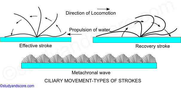
Effective stroke: During effective stroke, the cilium bends and beats against water thus bringing the body forward and sending the water backwards.
Recovery stroke: During recovery stroke, the cilium comes back to original position by its backward movement without any resistance.
Cilia shows two types of coordinated rhythms,
* Synchronous rhythm, where in the cilia beast simultaneously in a transverse row.
* Metachronous rhythm, where in cilia beat one after another in a longitudinal row. The metachronal waves pass from anterior to posterior end.
The beating of the cilia can be reversed to move backwards when a Paramecium encounters any undesirable object in its path. The ciliary movement is coordinated by infraciliary system though neuromotor center called as motorium present near the cytopharynx in the ciliates like Paramecium. The infraciliary system together with motorium form neuromotor system which helps in coordination of the beating of the cilia. Ciliary movement is the fastest locomotion in protozoans.
Paramecium feeds on holozoic manner and it is selective feeder. It feeds on bacteria, small protozoans, unicellular algae, diatoms etc. A single Paramecium can feed on 2-5 million bacteria in 24 hours. Feeding of the food is helped by the ciliary action. It feeds on the stationary or slowing swimming animals. The feeding apparatus of Paramecium are Buccal cavity, oral groove, vestibule and cytopharynx.
While feeding, the cilia of the oral groove beat strongly and thus it produces a current of water which brings in the food particles towards the vestibule. The cilia of the vestibule and buccal cavity pull in food particles. At the same time the unwanted food particles are rejected. The selected food is passed onto the cytopharynx through the route called as selection path. On the other hand the route through which the rejected food particles are sent out is called rejection path.
The ciliary membranelles i.e. peniculus and quadrulus lining the buccal cavity guide the selected food into the cytopharynx which remains separated from the endoplasm of the body by a membrane floor. The food collects here. The membrane enlarges and finally pinches off from the end of the cytopharynx as a food vacuole. Immediately a new food vacuole begins to form again.
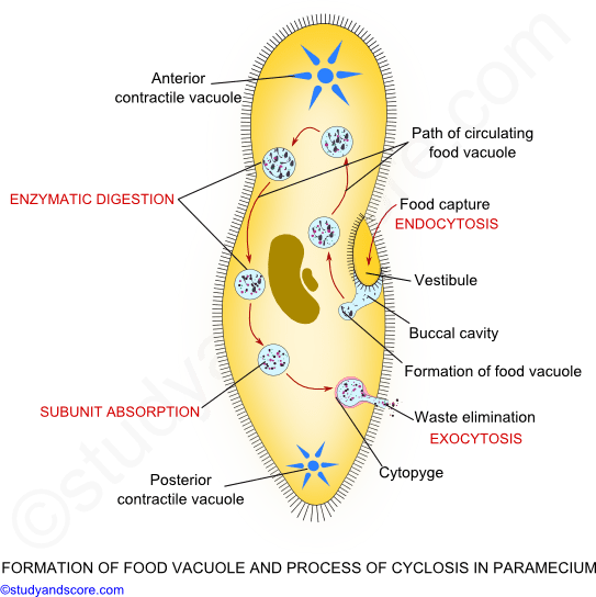
The food vacuoles are circulated around the body by the streaming endoplasm along a definite course. This streaming movement is called cyclosis. Several food vacuoles may be seen circulating in the endoplasm. The route of the food vacuole is as under:

The digestion of the ingested food occurs during cyclosis. Each of the food vacuole contains food particle and a little of water. The digestion is made by means of acids, alkalis and enzymes secreted from the surrounding endoplasm. Often mitochondria become associated with the food vacuoles to help in the digestion process. The food vacuole is initially acidic and in this acidic phase the prey is killed. The acid is hydro chloric acid. Later the vacuole becomes alkaline and most of the digestion occurs during this alkaline phase. Proteases, carbohydrates and lipases digest the food. The digested products are diffused into the endoplasm.

- Share with your friends! -
kaliyamoorthy Dass on Jul 07, 2018 at 01:20 pm
Dear Admin, your material is very useful for me but i cannot copy and download them. Can you please sent me all biology material. Please do needful. Thank you.


Study Score on Jul 07, 2018 at 06:25 pm
Thank you Kaliyamoorthy Dass. Our notes are copy protected. So you cannot copy or download them. However you can visit the site or make notes as per your need.
Login to post your comment here...
- or with social Account -