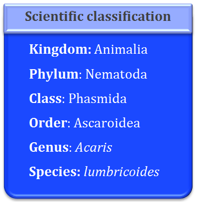
Nematodes are also known as roundworms. Ascaris lumbricoides is also known as the common roundworm. The roundworms are different from the flatworms and tapeworms as they have cylindrical body, pseudocoelom and a complete digestive tract lined by endodermal epithelium.
Most of these round worms are free living but some parasitic worms also exist. The species belonging to the genus Ascaris are large-sized and they inhabit in the intestines of the vertebrate hosts. Ascaris Lumbricoides is the most common roundworm which is the gastro-intestine parasite of the man.
Ascaris is cosmopolitan in distribution but it is chiefly found in India, China, Korea, Philippines and Pacific islands.
Ascaris is one of the most familiar endoparasites of humans. It has been reported in the intestines of pig, cattle, sheep, man and squirrels. It frequently inhabits the small intestine of children than adults.
Size and shape of the body: The body of the roundworms is elongated and cylindrical. It gradually tapers at both ends. The anterior end is more slender than that of the posterior end. The sex of the roundworms is separate with sexual dimorphism. The body of these worms is covered by cuticle, which has minute striations which imparts a pseudo segmented appearance to the worms.
Female roundworm measures about 20-40 cm in length and 4-6 mm in diameter, the posterior end of the female round worm is straight compared to that of the male. Male roundworms measure upto 20 cm in length and 2-4 mm in diameter. The males are smaller compared to the females. The posterior end of the male roundworms is curved.
Body color: The fresh specimens are light yellow to light pink in color. The semitransparency of the body wall enables visibility of some of the internal organs.
Longitudinal streaks: Along the entire length of the body of the roundworms, four longitudinal streaks are present namely one mid dorsal, one mid ventral and two lateral. The dorsal and ventral lines appear pure white whereas the lateral lines are more visible and appear brown in color. These longitudinal streaks are the impressions of the syncytial epidermis which is much thick along the median dorsal, median ventral and lateral positions.
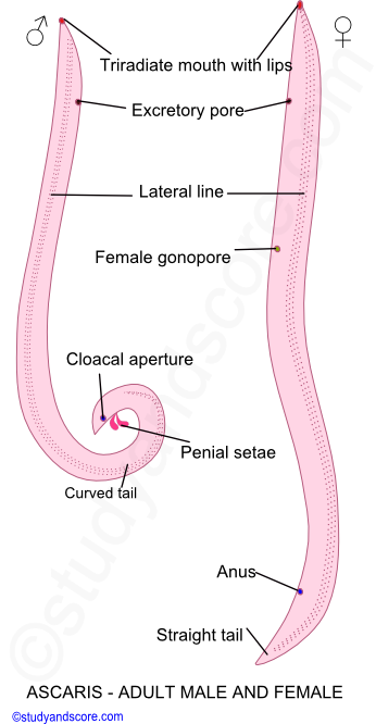
Anterior end: The anterior end of the roundworms is round and bears a triradiate mouth guarded by three broad lips or labia. One of the lips is situated mid-dorsally and the remaining two are situated sub-ventrally. The inner margin of each lip is forked, fleshy core made up of two anterior extensions of labial parenchyma and bears minute denticles. The outer surface of the lips bears minute sensory papillae.
Posterior end: Sexual dimorphism is clearly visible at the posterior end of these roundworms. The posterior end is straight in the female worms whereas it is curved in the male worms. In females, just before the tail ending, anus is present. This anus is guarded by lips. Only digestive tube opens outside through the anus. In males, anus is replaced by cloaca. Cloaca is the common aperture for the digestive tract ad genital tubes. Two chitinous spicules of equal size are seen protruding out of the cloacal aperture. These spicules are called as penial setae. These spicules help in the transfer of the sperms into the female vagina during the process of copulation. Also the tail end of the male Ascaris has numerous genital papillae which have a role in copulation.
Excretory pore: A single excretory pore lies mid-ventrally at a distance of about 2mm from the anterior end.
Female gonopore: The genital pore of the female lies mid-ventrally at about one-third distance from the anterior end. On the other hand the genital pore of the male opens into cloaca. The genital pore and anus open separately in the female roundworms.
The body wall of Ascaris comprises of three layers namely the outer cuticle, middle epidermis and inner longitudinal muscles.
The cuticle of these roundworms is thick, tough, transparent and glossy. This layer is secreted by the underlying epidermal cells. This is in continuation with the lining of the pharynx and rectum. In the young worms, the cuticle is shed off to permit the moulting and growth process.
The cuticle is permeable to salts, water and the products of metabolism. Also the enzymes present in the cuticle of Ascaris have the capability to control the passage of various metabolites. The cuticle also secretes anti-enzymes which have the neutralizing effect on the digestive juices of the host.
The cuticle in turn is divided into the following five layers:
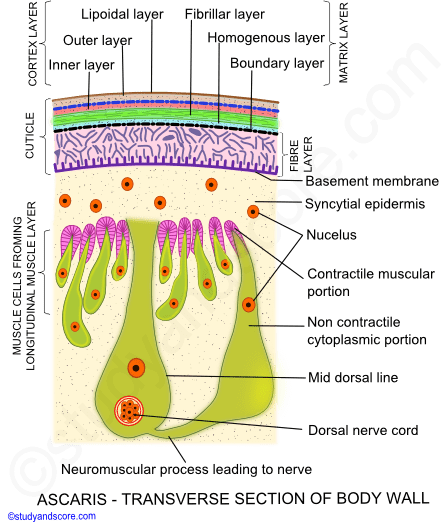
Epidermis is the syncytial layer present below the cuticle. The epidermis is thick along entire length of the body, at median dorsal, median ventral and two lateral positions where it forms the respective lateral lines. Epidermis is composed of relatively fewer cell. This layer has abundant reserves of fat and glycogen.
Circular muscles are absent in the round worms. The longitudinal muscles form single layer of spindle-shaped cells below the epidermis. The longitudinal muscles are interrupted at four places by longitudinal epidermal chord. The muscle layer is divided into four longitudinal columns and two ventro-lateral columns.
Each muscle cell has a fibrillar, contractile muscular portion and a granular non-contractile protoplasmic portion projecting into body cavity. The contractile muscular portion contains longitudinal contractile fibres arranged at intervals.
It also consists of the fibres for attachment to the cuticle. The non-contractile protoplasmic portion contains non-contractile supporting fibres, nucleus. The muscle tails of all the cells of the dorso-lateral columns are connected with the dorsal nerve cord whereas the muscle tails of all the cells of the ventro-lateral columns are connected with the ventral nerve cord.
A spacious fluid filled cavity is present between the body wall and the visceral organs. The fluid filled cavity is called as pseudo coelom and it cannot be considered as the true coelom as,
The pseudo coelom of Ascaris consists of five giant mesenchymal cells also known as pseudocoelomocytes. From one of these cells numerous cytoplasmic strands extend out in the form of fenestrated membranes. These membranes form delicate layers over visceral organs and muscles of the body wall.
The pseudo coelom is filled with odorous protein rich fluid called pseudo coelomic fluid. This fluid helps in transportation of metabolites and also keeps the body expanded. Other constituents of this fluid are glucose, nitrogenous substances, sodium chloride and phosphate.

- Share with your friends! -
Login to post your comment here...
- or with social Account -