Clitellum is the most important feature of reproduction in earthworm. Clitellum is secreted by specialized gland cells present in clitellar region. The clitellar region contains mucous cells, albumin cells and cocoon secreting cells. Mucous cells secrete mucous that forms the outer case of the cocoon. Albumin secreted by the albumin secreting cells is deposited along the zygotes in the cocoons. Cocoon is secreted by the cocoon secreting cells.
The earthworm is hermaphrodite or monoecious animals i.e. male and female reproductive organs are found in same individual. But in earthworms cross fertilization occurs because of the relative position of male and female genital apertures and protandrous nature of hermaphroditism.
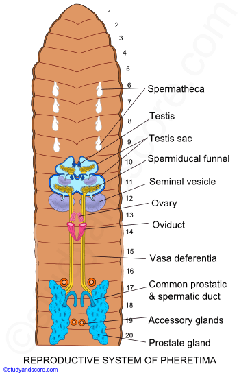
Male reproductive system
The male reproductive system in Pheretima consists of the following parts,
Testes: There are two pairs of testes. One pair is situated at 10th segment and another pair at 11th segment. These are situated ventrally beneath the stomach. Each pair is enclosed in the testis sac of that segment. They arise from the inner surface of anterior wall of the respective testis sac. Testes are small digitate bodies with 4 to 8 digitate processes and a compact base. They originate from the parietal coelomic epithelium. They produce spermatogonia or sperm mother cells.
Testes Sacs: Testes are enclosed by fluid filled sacs called testes sacs. One testes sac is found at 10th segment and other is at 11th segment. The testis sac of the 10th segment is small and it encloses testes and spermiducal funnels of that segment whereas the testis sac of 11th segment is comparatively large and it encloses testes, spermiducal funnels and seminal vesicle of that segment. On each side the testis sac communicates with a seminal vesicle through a small duct.
Seminal Vesicle: There are two pairs of seminal vesicles. One pair is situated at 11th and other at 12th segment. Each seminal vesicle communicates with testes sac. The spermatogonia are passed from testes into testes sacs and then they are passed into seminal vesicles where they develop into sperms. The mature sperms again go back into testes sac and pass into seminal funnel.
Spermiducal Funnel: There are two pairs of spermiducal funnel. One pair is found in 10th segment and another pair in 11th segment. They are ciliated and found below testes. Each funnel has fimbriated and ciliated margin
Vasa differentia: There are four tubes, which start running from 12th segment to 17th segment. In 17th segment, they unite with the duct of prostate gland to form common prostatic and spermatic duct. The vasa deferentia are internally lined by ciliated epithelium right up to the point, where they meet the common prostatic duct.
Prostate gland: There are two prostate glands. They are larger, irregular, flat, and wide. They extend from 16th or 17th segment to 20th to 21st segment. Each prostate gland has small curved duct, which joins with spermatic ducts and opens through male genital aperture at 18th segment.
Accessory gland: There are two pairs of accessory gland. One pair in 17th segment and another pair are in 19th segment. They are small and round in structure. They bear small ductules, which open through genital papillae.
Genital papillae: On the ventral side of 17th and 19th segments, there are two pairs of copulatory or genital papillae, one pair in each segment.
The spermatogonia produced in the testes are shed into the cavities of the testis-sacs from where they pass into the seminal vesicles. The spermatogonia undergo maturation into spermatozoa in the seminal vesicles. The mature sperms find their way back to testes sacs and pass out through the spermiducal funnels via vas differentia and male genital apertures.
Female reproductive system
The female reproductive system in Pheretima consists of the following parts,
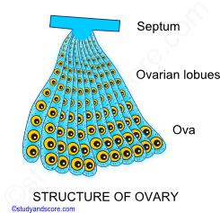
Ovaries: There is a pair of ovary situated at the 13th segment hanging on septum between 12th and 13th segment. Each ovary has finger like projections, which contained ova in series. The mature ova are found toward end and immature ova are found on the base of the lobules of the ovary.
Oviducal Funnel: One pair funnel like structures at 13th segment found below ovary with ciliated mouth called oviducal funnel. Each funnel opens into short conical tube called oviduct. Two oviducts fuse together at 14th segment and open outside through female genital aperture.
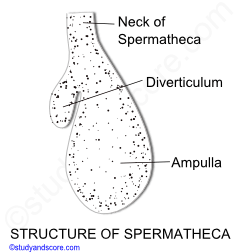
Spermathecae: There are four pairs of spermathecae. Each pair is found between 5-6, 6-7, 7-8, and 8-9th segment. Each spermatheca is flask shaped. The main body of spermatheca is called ampulla and short small lobe found attached at its side is called diverticulum. They store sperms.
Copulation
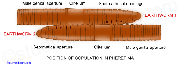
In rainy season, two earthworms copulate at night. During copulation, two earthworms get attached through their ventral surface with their anterior end pointing in opposite direction. The male genital aperture become erected and inserted into the spermathecal pore of each other. During the process sperms are exchanged together. Then they separate after about an hour. The sperms are received by spermathecae and are stored in their diverticula. The fertilization in earthworms is external and takes place in the cocoons.
Cocoon formation
After copulation, a membrane is secreted around clitellum by membrane secreting glands. The membrane starts to move towards anterior end of earthworm whereas the worm starts withdrawing itself backward. During the process, the membrane receives ova coming from female genital aperture and sperms coming from spermathecal pores. Lastly, the membrane is laid out on the ground. The elastic opening of membrane becomes closed. The structure is called cocoon (ootheca). Within cocoon, one of the sperm fertilizes the ovum to form zygote and young worm is developed inside the cocoon. The young worm comes out of cocoon after about 2-3 weeks. A dozen of cocoons are formed after copulation by each conjugant.
Development
The complete development from the fertilized egg up to the formation of young worms takes place within the cocoon. Though a cocoon contains many fertilized eggs, only one embryo develops. And this embryo grows at the expense of other eggs. All the other eggs become nursing cells and serve the growing embryo. Development is direct without any free larval stages. Cleavage is holoblastic, determinate, spiral and unequal resulting in the production of small micromeres and large megameres. Micromeres form the ectoderm and megameres form the endoderm.
The ovum is surrounded by a vitelline membrane and the cytoplasm is filled with yolk granules. The zygote is spherical in shape and undergoes cleavage. The micromeres are arranged in the animal hemisphere and the megameres are arranged in the vegetal hemisphere. As the cleavage proceeds, a hollow sphere or blastula is formed. The cavity of the blastula is called blastocoel or segmentation cavity.
The wall of the blastula is composed of a single layer of cells. The upper part of the blastula contains micromeres, while the lower part contains megameres. All the cells of the blastula continue to divide. Two large cells, which le side by side near the equator remain quiescent for some time. These are mesoblastic pole cells. They give rise to two rows of cells by successive divisions. These pole cells together with two rows of cell form mesoblastic bands. These mesoblastic bands form the future mesoderm.
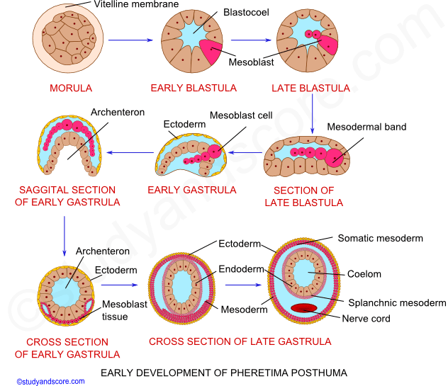
The spherical blastula becomes flattened and forms a solid oval structure with small cells on its upper surface and large columnar cells on its lower surface. The lower surface of the embryo starts to invaginate to form gastrula. As a result of this invagination, there appears an elongated groove on the lower surface. It leads into a cavity lined by original megameres. This cavity is referred to as archenteron and the elongated aperture is the blastopore. As the development proceeds, the lips of blastopore close from behind till only a small opening is left at the anterior end. This opening is the mouth. The tall ciliated cells of ectoderm that surround the mouth grow inwards to form stomodaeum. The stomadaeal canal leads into archenteron. On the formation of stomodaeum, the embryo starts feeding on the albumen present in the cocoon.
The two mesodermal bands lie parallel to each other. Each band divides in anterio-posterior axis into mesodermal somites. These somites are solid at first, but soon develop cavities within them. These are the coelomic cavities. These mesodermal somites grow upward and downward on either side between ectoderm and endoderm. Ultimately they fuse dorsally and ventrally. Thus outer layer is applied to the ectoderm to form somatopleure and inner layer is applied to the endoderm to form splachnopleure.
Somatic mesoderm forms the muscle of the body wall and the parietal peritoneum. Splanchnic mesoderm forms visceral peritoneum and muscles of alimentary canal. The transverse partitions between the somites form intersegmental septa. The ectodermal invagination of the last segment fuses with the enteron to form proctodaeum.
The primary embryonic layers five rise to the body structures. The ectoderm gives rise to epidermis, nervous system, stomodaeum, proctodaeum, excretory system and setal sacs. The mesoderm gives rise to muscles, coelomic epithelia and their derivatives, intersegmental septa, blood vessels and reproductive system. The endoderm gives rise to inner epithelial lining of alimentary canal and associated glands. Young earthworms when grown fully crawl out of the cocoon. The newly hatched young worm receives no parental care and they even resemble the adults except for size and absence of clitellum.

- Share with your friends! -
Login to post your comment here...
- or with social Account -