This parasite is cosmopolitan in distribution throughout all the sheep and cattle rearing areas of the world. It is of immense parasitological and pathological importance as it is the causative organism of Fascioliasis. Fascioliasis is a disease that causes great damage to the liver-tissues and the bile ducts of sheep and cattle.
Fasciola is digenetic endoparasites. It completes its life cycle in two hosts. Its primary host is sheep and secondary host is a freshwater gastropod either Limnea truncate or some species of Planorbis or Bulinus. Fasciola hepatica is also known as sheep liver fluke as it resides in the liver and bile ducts of sheep. It can also occur in other vertebrate hosts like goat, horse, dog, ass monkey, man , elephant etc. A single sheep may accommodate around 200 adult flukes in its liver and as a result the liver may stop to function. Consequently liver rot occurs.
Size and shape of the body: Tis parasite may measure about 1.8 to 3 cm long and 0.4 to 1.5 cm in width at the middle region. From the middle region the body tapers both anteriorly and posteriorly. Anterior end is a bit rounded whereas posterior end is pointed. It is a soft, oval, dorso-ventrally flattened leaf like animal.
Color of the body: The color of this animal is usually pinkish. The transparency of the body wall enables the view of internal organs like vitelline glands along the lateral margins, alimentary canals in the center.
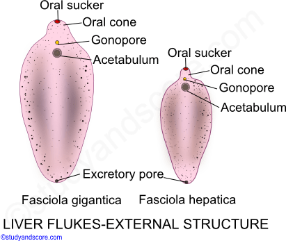
Oral cone: The anterior rounded end of this animal is prominently projected. This part is also called as oral cone or head lobe. At the tip of this oral cone a triangular aperture called mouth is located.
Suckers: This animal does not have any hooks or spines but has two small suckers namely, anterior sucker and the ventral sucker.
Apertures: Along with mouth there are two more apertures namely gonopore and excretory pore. The gonopore is a small aperture lying in front of the acetabulum. A single excretory pore is situated at the posterior end of the body. Because of the presence of an incomplete alimentary canal, there is no anus in this animal.
Scales: The body of Fasciola is covered by tegument from which numerous minute backwardly directed spinules or scales arise. These spinules help in locomotion, protection and in anchorage of the parasite body in the bile ducts of the host.
The body wall of Fasciola consists of the following parts,
It is the external covering of the body. Tegument is thick and non-ciliated structure containing mitochondria, endoplasmic strands, vacuoles and pinocytotic vesicles. This tegument layers helps the animal to overcome the digestive action of the host fluids. Also tegument bears spinules which in turn help in locomotion, protection and in anchorage of the parasite body in the bile ducts of the host.
Just under the tegument, a thin and well defined acidophilic basement membrane is present.
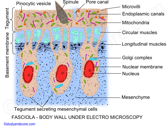
Fasciola has three muscle layers namely circular muscle layer, longitudinal muscle layer and diagonal muscle layer. Bothe circular and longitudinal muscles layers together are called as integumentary muscles. On the other hand diagonal muscles are present inside the longitudinal muscle layer. All these muscle fibers are smooth type. In the suckers, these muscles form a stout bundle like structure called as radial fiber which imparts radial striations to them.
This layer is of mesodermal origin. All the internal organs are embedded in it and thus it acts as a packing material between the internal organs. The mesenchyme consists of irregular uninucleate and binucleated cells with large fluid-filled intercellular spaces. Some of these cells secrete the tegument layer. Along with the skeletal function, mesenchyme also helps in transport.
Fasciola has an incomplete alimentary canal and thus it do not have anus.
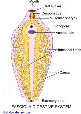
The flukes when hungry migrate into smaller bile ducts and capillaries for feeding. They suck the lymph, bile and tissue pieces with the help of the oral sucker. The digestion in these flukes is extracellular type and it takes place in the intestine.
The distribution of the digested food is done by diverticula of the intestine. Monosaccharides can directly diffuse through the body surface. Amino acids are absorbed by the tegument. The food is reserved in the form of glycogen and fats in the mesenchyme and muscles.
The excretory system of Fasciola is also known as Protonephredial system. It consists of following parts,
The flame cells are modified mesenchymal cells to perform the excretory function. Each flame cell consists of intracellular lumen in which few long cilia are present. Each of these cilia arises from a basal granule. As these structures vibrate like a flickering of a flame they are named so.
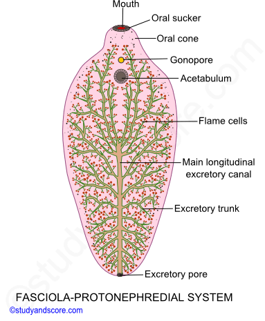
The lumen of the flame cell is in continuation with the microscopic excretory or capillary ducts. The capillary ducts from a few protonephridia open into narrow collecting tubule. Several such tubules open into larger twigs which in turn open into vessels. The excretory vessels of the anterior part of body open into four trunks.
These four trunks unite posteriorly to form a single longitudinal excretory canal. This longitudinal excretory canal extends up to posterior end of the body where it opens out through single excretory pore. The excretory vessels of the posterior part, open directly into the longitudinal excretory canal. All the ducts except the median longitudinal canal are lined with cilia.
As flukes are parasitic in nature, they do not have sense organs. They possess developed nervous system. Brain forms a cerebral ring around pharynx and it bears a pair of lateral cerebral ganglia and ventral ganglia. Fine nerves arise from the brain and supply the anterior and posterior regions.
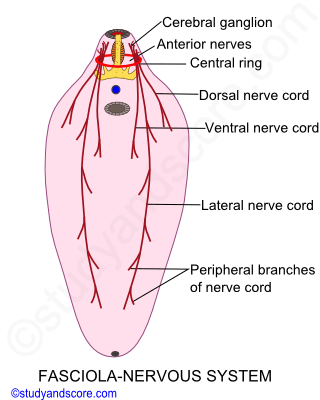
The suckers receive more supply. Three pairs of longitudinal nerve cords extend posteriorly and they give out numerous peripheral branches to various organs. Of these three pairs one is dorsal; one ventral and last one is lateral. Out of these three lateral cords are well developed and are connected by few transverse commissures.

- Share with your friends! -
Login to post your comment here...
- or with social Account -